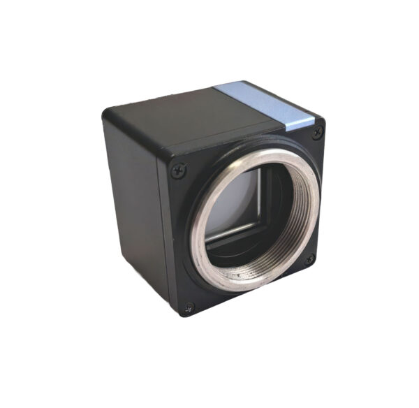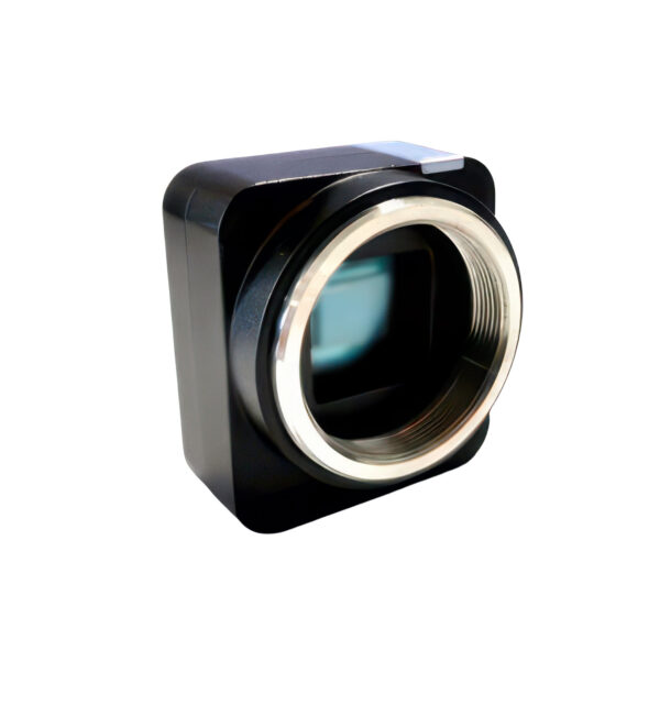
Wavefront camera for precise phase and intensity measurement
The MAKATEA Cam from Silios is a compact wavefront imaging camera for quantitative phase imaging (QPI) in scientific and industrial applications.
Through the QLSI technology (Quadriwave Lateral Shearing Interferometry) the system records Intensity and phase information simultaneously and thus enables a highly sensitive analysis of optical wavefronts.
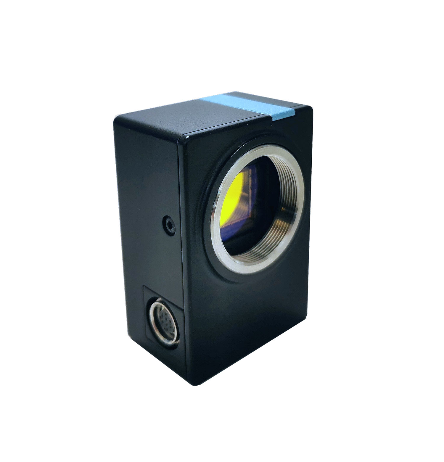
The integrated micro-optical grating structure in front of the CMOS sensor allows the direct reconstruction of the wavefront and the simultaneous recording of an intensity image. This allows Quantitatively characterize sample structures and optical systems.
The camera works in visible spectral range from 400-900 nm and achieves a Phase sensitivity of < 1 nm RMS. This makes the system particularly suitable for applications in which very small optical changes need to be detected.
Typical areas of application are
-
Quantitative phase microscopy
-
label-free cell analysis
-
Nanophotonics
-
Materials research
-
Optical wavefront analysis
Thanks to the compact design and the C-/CS-mount interface the camera can be easily integrated into existing Microscopy or measuring systems integrate. Data is transmitted via USB 3.0, optional a External trigger is available for synchronized measurements.
For extended applications, the WaveFront LAB SDK available. This allows Raw data, wavefront maps, gradient and transmission images visualized and processed in real time.
Comparison of the Silios wavefront cameras:
| Model | spectral range | Main strength | Sensor resolution | Wavefront resolution | Technology | Typical applications |
|---|---|---|---|---|---|---|
| MAKATEA Cam | 400-900 nm (visible) | Very high phase sensitivity (< 1 nm RMS) | 2048 x 2048 (4.2 MP) | 680 x 680 | QLSI (Quadriwave Lateral Shearing Interferometry) | Quantitative phase microscopy, cell analysis, label-free microscopy |
| TIKEI Cam | 400-900 nm (visible) | High spatial resolution for wavefront analysis | 4500 x 4500 ( 20 MP) | 1500 x 1500 | Micro-optical wavefront structure | Microscopy, material analysis, optical system diagnostics |
| TAKUME SWIR Cam | 900-1700 nm (SWIR) | Wavefront measurement in the infrared range | 1296 × 1032 (1.3 MP) | 432 × 344 | COLOR-SHADES® technology | Metasurface measurements, nanoparticle analysis, optical metrology |